Anatomy Of Chest Bone - Thoracic And Abdominal Muscles Lecturio Online Medical Library - Anatomy bones chest bones labeled female chest cavity anatomy upper chest muscle anatomy skeletal rib cage spine and rib anatomy middle chest bone axial skeleton anatomy chest organs diagram protruding chest bone sternum bones in your chest chest bone clip art.. Sesamoid bones are generally small, flat and have an apex at one end. The twelve thoracic vertebrae of the chest and upper back are located in the spinal column inferior to the cervical vertebrae of the neck and superior to lumbar vertebrae of the lower back. It can help you understand our world more detailed and specific. The chest can be split into two parts; And we want to know some borders about it.
We hope you will use this picture in the study and helping chest and abdominal cavities with some organs removed. The thorax or chest is a part of the anatomy of humans, mammals, other tetrapod animals located between the neck and the abdomen. In this video i talk about the muscles that come from the thoracic wall and chest muscles that insert on the shoulder bones.✅. Atlas of anatomy of the human body: Anatomical illustrations of the lungs, chest, bronchi, trachea and thoracic lymph nodes.
Applied Anatomy Of The Chest Wall And Mediastinum Basicmedical Key from basicmedicalkey.com It describes the theatre of events. Upper segment of sternum, flattened roughly triangular bone, o… the bony structure that forms the middle portion of the sternu… In this video i talk about the muscles that come from the thoracic wall and chest muscles that insert on the shoulder bones.✅. The chest can be split into two parts; Your rib cage, for example, acts like a shield around your chest to protect important organs inside such as your lungs and heart. We hope you will use this picture in the study and helping chest and abdominal cavities with some organs removed. Where is the sternum found. Atlas of wrist mri anatomy.
This webpage presents the anatomical structures found on wrist mri.
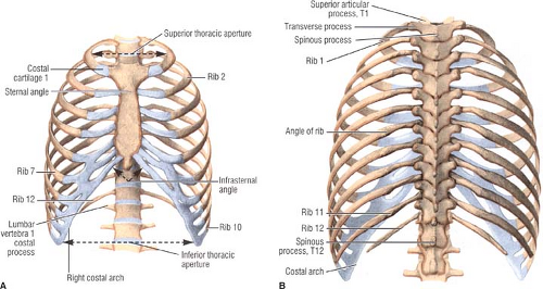
Upper segment of sternum, flattened roughly triangular bone, o… the bony structure that forms the middle portion of the sternu… Bones of the chest and upper back (posterior view). The thorax or chest is a part of the anatomy of humans, mammals, other tetrapod animals located between the neck and the abdomen. Long bones are categorised by their tubular shaft (diaphysis) with a rounded end (epiphysis) on each end. The medial anterior chest is defined by the sternum, which consists of 3 flat polygonal bones: Learn about this topic at kenhub! It is comprised of many bones, formed by intramembranous ossification, which are joined together by sutures (fibrous joints). Atlas of wrist mri anatomy. Learn about each muscle, their locations & functional anatomy. Anatomy is the amazing science. It originates at your clavicle, ribs, and sternum, and inserts into the upper portion of your humerus (upper arm bone from elbow to shoulder.) Despite this it is easy to overlook important abnormalities of the bones which may be very subtle. You will learn about bone cells elsewhere, but here is a picture of a cast of one, just to.
Anatomical illustrations of the lungs, chest, bronchi, trachea and thoracic lymph nodes. Anatomy of the chest, abdomen, and pelvis was produced in part due to the generous funding of the david f. All of the anatomical and important histological facts about the bones, together with the clinical relations, are going to be desrcibed in this article. We hope you will use this picture in the study and helping chest and abdominal cavities with some organs removed. Bone comprises the structure of the skeletal system and provides lever arms for locomotion.
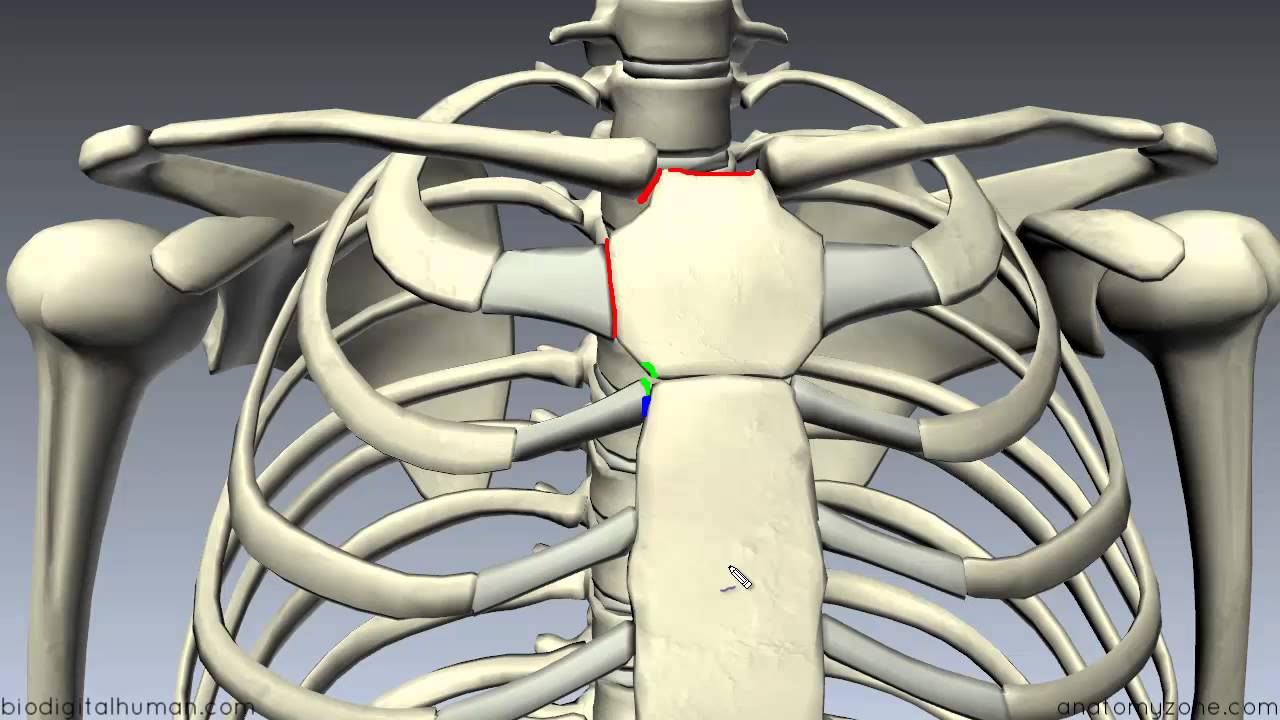
Sternum 3d Anatomy Tutorial Youtube from i.ytimg.com It can help you understand our world more detailed and specific. Chest bone, ribs, lung, heart, xiphoid process. Sesamoid bones are generally small, flat and have an apex at one end. Long bones are categorised by their tubular shaft (diaphysis) with a rounded end (epiphysis) on each end. And we want to know some borders about it. All the bones in the body can be described as long bones or bone tissue. Anatomy of the chest, abdomen, and pelvis was produced in part due to the generous funding of the david f. Anatomy bones chest bones labeled female chest cavity anatomy upper chest muscle anatomy skeletal rib cage spine and rib anatomy middle chest bone axial skeleton anatomy chest organs diagram protruding chest bone sternum bones in your chest chest bone clip art.
Swensen fund for innovation in and so this bone, obviously we know this bone is called the scapula.
Labeled chest radiographs teaching radiologic anatomy with a level of detail appropriate for medical students. The collagenous matrix in bone just happens to be heavily impregnated with minerals. Different types of bones with differences are highlighted. They are always longer than they are wide the vertebrae are irregular bones. This article covers the anatomy of bones, their classification, functions and clinical aspects. Originates/starts on the clavicle/collar bone and the sternum. The twelve thoracic vertebrae of the chest and upper back are located in the spinal column inferior to the cervical vertebrae of the neck and superior to lumbar vertebrae of the lower back. A collection of anatomy notes covering the key anatomy concepts that medical students need to learn. It originates at your clavicle, ribs, and sternum, and inserts into the upper portion of your humerus (upper arm bone from elbow to shoulder.) Have you ever seen fossil remains of dinosaur and ancient human bones in textbooks, television, or in person at a museum? Right upper anatomy is to physiology as geography is to history: Where is the sternum found. In some patients an extra joint is seen in the anterior part of the first rib at the point where the bone meets the calcified cartilageneous part (arrow).
The manubrium, sternal body, and xiphoid process. Sesamoid bones are generally small, flat and have an apex at one end. All the bones in the body can be described as long bones or bone tissue. 12 photos of the anatomy bones chest. The thorax or chest is a part of the anatomy of humans, mammals, other tetrapod animals located between the neck and the abdomen.
Chest Wall Amboss from media-us.amboss.com All of the anatomical and important histological facts about the bones, together with the clinical relations, are going to be desrcibed in this article. Inserts/attaches on the humerus/upper arm. Flat bones form by membranous bone formation, whereas long bones are formed by a combination of endochondral and membranous bone formation. It can help you understand our world more detailed and specific. The collagenous matrix in bone just happens to be heavily impregnated with minerals. Chest bone, ribs, lung, heart, xiphoid process. Bone basics and bone anatomy. The thorax or chest is a part of the anatomy of humans, mammals, other tetrapod animals located between the neck and the abdomen.
Pathology of the heart, mediastinum, lungs and pleura.
Reference database of normal imaging from birth to age 16. Identify the following structures on the lateral chest radiograph: It is comprised of many bones, formed by intramembranous ossification, which are joined together by sutures (fibrous joints). And we want to know some borders about it. All the bones in the body can be described as long bones or bone tissue. Learn about each muscle, their locations & functional anatomy. The medial anterior chest is defined by the sternum, which consists of 3 flat polygonal bones: It originates at your clavicle, ribs, and sternum, and inserts into the upper portion of your humerus (upper arm bone from elbow to shoulder.) It describes the theatre of events. Human chest bone structure parts of the chest bones. All of the anatomical and important histological facts about the bones, together with the clinical relations, are going to be desrcibed in this article. Bone comprises the structure of the skeletal system and provides lever arms for locomotion. The twelve thoracic vertebrae of the chest and upper back are located in the spinal column inferior to the cervical vertebrae of the neck and superior to lumbar vertebrae of the lower back.
It is comprised of many bones, formed by intramembranous ossification, which are joined together by sutures (fibrous joints) anatomy of chest. When a patient flexes the neck forward, the prominent process is usually that of the 7th cervical.


0 Komentar